Persistent Trigeminal Artery
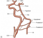
The persistent carotid-vertebrobasilar anastomoses are variant anatomical arterial communications between the anterior and posterior circulations due to abnormal embryological development of the vertebrobasilar system. Four notable caroticobasilar connections that may persist into adult life are the trigeminal, otic, hypoglossal, and proatalantal intersegmental arteries.
Although discovered incidentally, an altered hemodynamic may lead to an increased association of aneurysms, vascular malformations, and stroke. It is extremely critical to aware of the presence and significance of embryonic arterial communications when interpreting imaging and planning interventions.

Figure 1. Schematic diagram showing the series of embryonic anastomoses between the anterior and posterior circulation which form and regress until the final one, the posterior communicating artery, is established. [1]
Background
Persistent trigeminal artery (PTA) was first described in cadaveric brain by Richard Quinn in 1844, but it was only in 1950 with the help of angiography that Sutton reported its presence in live patients. Persistent trigeminal artery (PTA) is the most cephalic and the most frequent of the carotid-basilar anastomosis, comprising up to 85% of all communications, with an angiographic incidence between 0.1%–0.6%. [2], [3],
Classification
Numerous classification systems have been developed to describe the PTA. Saltzman et al. [7] classified the angiographic appearance of PTA by conventional angiography into three types according to their relationship with the neighboring vessels. Weon et al. [8] proposed a new classification scheme for PTA based on magnetic resonance (MR) angiography, identifying five different types based on their anatomical relationship to the neighboring arteries. Salas et al. [9] classified PTA into two different types depending on the relationship with the abducens nerve.[10]

Figure 2. (A) Saltzman 1 (Fetal type)/Weon I, (B) Saltzman 2 (Adult type)/Weon II, (C) Saltzman combined type (1+2)/Weon III, (D) Saltzman combined type (1+2)/Weon IV, (E) Saltzman type 3a/Weon Va, (F) Saltzman type 3b/Weon Vb, and (G) Saltzman type 3c/Weon Vc. Red arrows indicate blood supply by the persistent trigeminal artery (PTA). Blue arrows indicate blood supply by the posterior communicating artery (PComA). Dashed lines indicate hypoplastic arteries, reduced or absent (in the case of posterior communicating artery) flow between the segment. AICA, anterior inferior cerebellar artery; BA, basilar artery; PCA, posterior cerebral artery; PICA, posterior inferior cerebellar artery; SCA, superior cerebellar artery; VA, vertebral artery.[5]
Clinical features
Usually PTAs are incidentally identified. Rarely, trigeminal neuralgia has been reported.
Characteristic Radiologic Findings
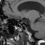
A characteristic tau sign or trident sign is described as its appearance on sagittal CTA or MR images [6]. The tau sign represents the appearance of a persistent trigeminal artery on the sagittal plane of an angiogram or on sagittal MRI images. It resembles the Greek letter τ. The trident sign of a persistent trigeminal artery refers to the appearance of the intracranial circulation on lateral projection.

Figure 3. Sagittal T1WI demonstrates an abnormal configuration of vessels in the presellar ICA. The combination of the vertical and horizontal segments of the ICA and the proximal persistent trigeminal artery (outlined in white) creates the outline of the Greek letter τ, described as the tau sign. [4]

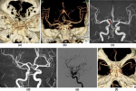
Figure 4. Type 1 persistent trigeminal artery (PTA) a supplies both posterior cerebral arteries (PCAs) and both superior cerebellar arteries (SCAs). The proximal portion of the basilar artery (BA) connected with the PTA shows moderate hypoplasia (arrow PTA). Type 2 PTA b supplies both SCAs. Both posterior communicating arteries (PComA) supply both PCAs (red arrow). Proximal portion of the BA connected with the PTA demonstrates severe hypoplasia (black arrow PTA). Type 3 PTA (c and d in the same patient, yellow arrow PTA) supplies the contralateral PCA (red arrow contralateral PCA) and the ipsilateral PComA supplies the ipsilateral PCA (white arrow ipsilateral PCA). Type 4 PTA (e and f in the same patient). Right internal carotid angiography (e) shows trigeminal artery supplying ipsilateral PCA. Contralateral PComA (white arrow contralateral PComA) supplies the PCA and proximal portion of BA connected with the PTA shows no hypoplasia (f) [4]

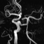
Figure 5. 3D TOF MRA MIP image demonstrates the persistent trigeminal artery. Notice the relative hypoplasia of the proximal basilar artery; also, note absence of the posterior communicating artery. [4]

Figure 6. (A) T2-weighted axial image showing a transsellar or medial course of the left type I persistent trigeminal artery ( red arrows ). (B) Time-of-flight image of the same patient. (C) Sagittal magnetic resonance angiography image showing the course of the persistent trigeminal artery ( red arrows ) and the hypoplastic basilar artery ( white arrows ). [5]
Related pathology
Leading to an increased association of aneurysms, vascular malformations, and stroke because of an altered hemodynamic.
*References
1. Christen D. Barras , “Current Status of Imaging of the Brain and Anatomical Features”, Grainger & Allison's Diagnostic Radiology 2021, 7th, 53, 1351-1386
2. Alcalá-Cerra G., Tubbs R.S., and Niño-Hernández L.M.: Anatomical features and clinical relevance of a persistent trigeminal artery. Surg Neurol Int 2012; 3: pp. 111
3. Azab W., Delashaw J., and Mohammed M.: Persistent primitive trigeminal artery: a review. Turk Neurosurg 2012; 22: pp. 399-406
4. Girish M. Fatterpekar MD, “Case 68”, Teaching Files: Brain and Spine 2012, The, 136-137
5. Gaurav Tyagi, “Persistent Trigeminal Artery: Neuroanatomic and Clinical Relevance” World Neurosurgery, 2020, Volume 134, Pages e214-e223.
6. Goyal M. The tau sign. (2001) Radiology. 220 (3): 618-9.
7. Saltzman GF (1959) Patent primitive trigeminal artery studied by cerebral angiography. Acta Radiol 51:329–336
8. Weon YC, Choi SH, Hwang JC, Shin SH, Kwon WJ, Kang BS (2011) Classification of persistent primitive trigeminal artery (PPTA): a reconsideration based on MRA. Acta Radiol 52:1043–1051
9. Salas E, Ziyal IM, Sekhar LN, Wright DC (1998) Persistent trigeminal artery: an anatomic study. Neurosurgery 43:557–562
10. Myeong Jin Kim and Myoung Sao Kim, Persistent primitive trigeminal artery: analysis of anatomical characteristics and clinical significances, "Surgical and Radiologic Anatomy" volume 37, pages 69–74 (2015)
The persistent carotid-vertebrobasilar anastomoses are variant anatomical arterial communications between the anterior and posterior circulations due to abnormal embryological development of the vertebrobasilar system. Four notable caroticobasilar connections that may persist into adult life are the trigeminal, otic, hypoglossal, and proatalantal intersegmental arteries.
Although discovered incidentally, an altered hemodynamic may lead to an increased association of aneurysms, vascular malformations, and stroke. It is extremely critical to aware of the presence and significance of embryonic arterial communications when interpreting imaging and planning interventions.
- The persistent trigeminal artery (PTA): arises from proximal cavernous internal carotid artery (ICA)
- The primitive hypoglossal artery (PHA): arises from cervical ICA at C1 to C3 levels
- The persistent otic artery (POA): arises from petrous ICA
- The proatlantal intersegmental artery (PIA): arises from internal carotid artery or from external carotid artery

Figure 1. Schematic diagram showing the series of embryonic anastomoses between the anterior and posterior circulation which form and regress until the final one, the posterior communicating artery, is established. [1]
Background
Persistent trigeminal artery (PTA) was first described in cadaveric brain by Richard Quinn in 1844, but it was only in 1950 with the help of angiography that Sutton reported its presence in live patients. Persistent trigeminal artery (PTA) is the most cephalic and the most frequent of the carotid-basilar anastomosis, comprising up to 85% of all communications, with an angiographic incidence between 0.1%–0.6%. [2], [3],
Classification
Numerous classification systems have been developed to describe the PTA. Saltzman et al. [7] classified the angiographic appearance of PTA by conventional angiography into three types according to their relationship with the neighboring vessels. Weon et al. [8] proposed a new classification scheme for PTA based on magnetic resonance (MR) angiography, identifying five different types based on their anatomical relationship to the neighboring arteries. Salas et al. [9] classified PTA into two different types depending on the relationship with the abducens nerve.[10]

Figure 2. (A) Saltzman 1 (Fetal type)/Weon I, (B) Saltzman 2 (Adult type)/Weon II, (C) Saltzman combined type (1+2)/Weon III, (D) Saltzman combined type (1+2)/Weon IV, (E) Saltzman type 3a/Weon Va, (F) Saltzman type 3b/Weon Vb, and (G) Saltzman type 3c/Weon Vc. Red arrows indicate blood supply by the persistent trigeminal artery (PTA). Blue arrows indicate blood supply by the posterior communicating artery (PComA). Dashed lines indicate hypoplastic arteries, reduced or absent (in the case of posterior communicating artery) flow between the segment. AICA, anterior inferior cerebellar artery; BA, basilar artery; PCA, posterior cerebral artery; PICA, posterior inferior cerebellar artery; SCA, superior cerebellar artery; VA, vertebral artery.[5]
Clinical features
Usually PTAs are incidentally identified. Rarely, trigeminal neuralgia has been reported.
Characteristic Radiologic Findings
A characteristic tau sign or trident sign is described as its appearance on sagittal CTA or MR images [6]. The tau sign represents the appearance of a persistent trigeminal artery on the sagittal plane of an angiogram or on sagittal MRI images. It resembles the Greek letter τ. The trident sign of a persistent trigeminal artery refers to the appearance of the intracranial circulation on lateral projection.

Figure 3. Sagittal T1WI demonstrates an abnormal configuration of vessels in the presellar ICA. The combination of the vertical and horizontal segments of the ICA and the proximal persistent trigeminal artery (outlined in white) creates the outline of the Greek letter τ, described as the tau sign. [4]

Figure 4. Type 1 persistent trigeminal artery (PTA) a supplies both posterior cerebral arteries (PCAs) and both superior cerebellar arteries (SCAs). The proximal portion of the basilar artery (BA) connected with the PTA shows moderate hypoplasia (arrow PTA). Type 2 PTA b supplies both SCAs. Both posterior communicating arteries (PComA) supply both PCAs (red arrow). Proximal portion of the BA connected with the PTA demonstrates severe hypoplasia (black arrow PTA). Type 3 PTA (c and d in the same patient, yellow arrow PTA) supplies the contralateral PCA (red arrow contralateral PCA) and the ipsilateral PComA supplies the ipsilateral PCA (white arrow ipsilateral PCA). Type 4 PTA (e and f in the same patient). Right internal carotid angiography (e) shows trigeminal artery supplying ipsilateral PCA. Contralateral PComA (white arrow contralateral PComA) supplies the PCA and proximal portion of BA connected with the PTA shows no hypoplasia (f) [4]

Figure 5. 3D TOF MRA MIP image demonstrates the persistent trigeminal artery. Notice the relative hypoplasia of the proximal basilar artery; also, note absence of the posterior communicating artery. [4]

Figure 6. (A) T2-weighted axial image showing a transsellar or medial course of the left type I persistent trigeminal artery ( red arrows ). (B) Time-of-flight image of the same patient. (C) Sagittal magnetic resonance angiography image showing the course of the persistent trigeminal artery ( red arrows ) and the hypoplastic basilar artery ( white arrows ). [5]
Related pathology
Leading to an increased association of aneurysms, vascular malformations, and stroke because of an altered hemodynamic.
*References
1. Christen D. Barras , “Current Status of Imaging of the Brain and Anatomical Features”, Grainger & Allison's Diagnostic Radiology 2021, 7th, 53, 1351-1386
2. Alcalá-Cerra G., Tubbs R.S., and Niño-Hernández L.M.: Anatomical features and clinical relevance of a persistent trigeminal artery. Surg Neurol Int 2012; 3: pp. 111
3. Azab W., Delashaw J., and Mohammed M.: Persistent primitive trigeminal artery: a review. Turk Neurosurg 2012; 22: pp. 399-406
4. Girish M. Fatterpekar MD, “Case 68”, Teaching Files: Brain and Spine 2012, The, 136-137
5. Gaurav Tyagi, “Persistent Trigeminal Artery: Neuroanatomic and Clinical Relevance” World Neurosurgery, 2020, Volume 134, Pages e214-e223.
6. Goyal M. The tau sign. (2001) Radiology. 220 (3): 618-9.
7. Saltzman GF (1959) Patent primitive trigeminal artery studied by cerebral angiography. Acta Radiol 51:329–336
8. Weon YC, Choi SH, Hwang JC, Shin SH, Kwon WJ, Kang BS (2011) Classification of persistent primitive trigeminal artery (PPTA): a reconsideration based on MRA. Acta Radiol 52:1043–1051
9. Salas E, Ziyal IM, Sekhar LN, Wright DC (1998) Persistent trigeminal artery: an anatomic study. Neurosurgery 43:557–562
10. Myeong Jin Kim and Myoung Sao Kim, Persistent primitive trigeminal artery: analysis of anatomical characteristics and clinical significances, "Surgical and Radiologic Anatomy" volume 37, pages 69–74 (2015)
