Lac Phan
New member
- Messages
- 12
- Reaction score
- 10
Case study
ARTERIOVENOUS MALFORMATION
1/ Patient name: T.H.T 16 yo, Male
Adress: Thanh Binh – Dong Thap
Admission date: 06:13.pm 25 th of June 2021
2/ Chief complaint: peripheral facial palsy
3/ History of present illness: The disease started with the sundenly onset of peripheral facial palsy, no treatment, unremitting à admitted.
Clinical examination:
Vital sign: Pulse 102 bpm, Temp 37oC, BP 120/70 mmHg, RR 20 bpm
Neurologic exams: GCS 15 points, pupil reflex (+) on both sides, no paralysis in both side, peripheral facial palsy on the left side.
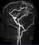
MRI image

An AVM in the right occipital lobe, size about 18x35mm

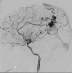
The AVM is located in right occipital lobe, the size is about 18x35mm. It is supplied by the anterior, posterior and middle cerebral arteries, which are drained to the superior sagittal sinus.

Before Embolization

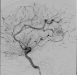
After Embolization by glue

CT-scan imagine after embolization: high density injury in the Parietal lobe
After 5 days of treament in S.I.S hospital, the patient has a full recovery, with no focal neurological signs.
ARTERIOVENOUS MALFORMATION
1/ Patient name: T.H.T 16 yo, Male
Adress: Thanh Binh – Dong Thap
Admission date: 06:13.pm 25 th of June 2021
2/ Chief complaint: peripheral facial palsy
3/ History of present illness: The disease started with the sundenly onset of peripheral facial palsy, no treatment, unremitting à admitted.
Clinical examination:
Vital sign: Pulse 102 bpm, Temp 37oC, BP 120/70 mmHg, RR 20 bpm
Neurologic exams: GCS 15 points, pupil reflex (+) on both sides, no paralysis in both side, peripheral facial palsy on the left side.
MRI image

An AVM in the right occipital lobe, size about 18x35mm

The AVM is located in right occipital lobe, the size is about 18x35mm. It is supplied by the anterior, posterior and middle cerebral arteries, which are drained to the superior sagittal sinus.

Before Embolization

After Embolization by glue

CT-scan imagine after embolization: high density injury in the Parietal lobe
After 5 days of treament in S.I.S hospital, the patient has a full recovery, with no focal neurological signs.
