Ginonguyen
New member
- Messages
- 4
- Reaction score
- 1
A. Administration
- Name: NGUYEN HOANG A
- Date of birth: 07/06/1977
- Hospitalized day: 27/01/2021
- Reason for hospitalization: Headache, swollen and painful right eye.
B. Antecedent:
- Hypertension for 2 years.
- No history of head injury.
C. Medical history:
The patient has had a headache for more than 6 months now, treatment in many places does not reduce. Accompanying symptoms are ringing in the ears, swelling and pain in the right eye, and red eyes. Medications the patient used in the past: Pain relievers, eye drops for conjunctivitis, antibiotics, anti-inflammatory drugs, tinnitus medications.
However, the above unpleasant symptoms did not subside, but gradually worsened. At the same time, they make him lose sleep, worry, affect his daily work. Therefore, he decided to go to the Can Tho Heart and Stroke Hospital. Here, he was examined by doctors and diagnosed with Carotid Cavernous fistula disease.
D. Subclinical
This patient was assigned blood tests and imaging tests. After that, the patient underwent a DSA and was diagnosed with: Carotid Cavernous fistula disease type A.

E. Treatment results
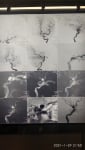
The following is a report of the DSA scan and treatment for Carotid Cavernous fistula disease type A at this patient:


And the results of the DSA check after treatment are as follows:

F. Postoperative developments
After 2 days of treatment, he felt relief from headaches, ringing in his ears and swelling in his right eye. And he was discharged from the hospital 1 day later
G. Disscussion
Carotid cavernous fistulas (CCFs) are rare, abnormal connections between the carotid artery and cavernous sinus, which can be either direct (high flow) or indirect (low flow) communication. Barrow et al. reported a classification of CCFs as follows: direct (high-flow) communication of the carotid artery and cavernous sinus (Type A), indirect communications of the cavernous sinus with branches of the cavernous carotid artery (Type B), or dural branches of external carotid artery (Type C), or both the branches of external and internal carotid arteries (Type D).
The CCFs may occur spontaneously or following secondary causes (trauma, vascular aneurysm or malformations and venous thrombosis). Direct CCFs are often caused by head trauma or rupture of intra-cavernous aneurysm or head surgery.
There is limited data regarding the incidence of CCFs, either primary or secondary types. However, through retrospective analysis, Liang et al. reported that the overall incidence of traumatic CCF was 3.8% among patients with basilar skull fractures. Sudden increase in pressure within the internal carotid artery (ICA) may be the mechanism for the development of direct CCF. Based on the different types, the presentation of CCFs is highly variable. Direct CCFs mostly present with orbital bruit (80%), proptosis (72%), chemosis (55%), abducens nerve palsy (49%), and conjunctival injection (44%). Vision loss is one of the most fearful complications of CCFs. Sight threatening complications may occur in direct CCF due to severe exposure keratopathy, corneal ulcerations, and possibly central retinal artery occlusion. Currently, no data is available for incidence of vision loss if the direct CCF is not properly and timely treated.
Indirect CCFs often occur spontaneously and are more common in older women. The cause of indirect CCFs is unknown and their presentations can vary widely by the patterns of venous drainage and the speed of blood flow. Indirect CCF especially of anterior drainage pattern may threaten the vision through increased intraocular pressure (due to venous congestion).

The differential diagnoses of CCFs are cerebral aneurysms, vascular malformations of eyes, inflammation of orbits, retro-orbital cellulitis, thyroid exophthalmos, retrobulbar hemorrhage, tumor of lacrimal gland, cavernous sinus thrombosis, and vasculitis. CCFs may be suggested by clinical evaluation and imaging studies, especially MR-orbits and CTA or MRA of the head. However, the gold standard test for CCFs remains carotid digital arteriography [5]. Treatment decision depends on the severity of clinical presentations. Generally, treatment of CCFs is suggested if there are worsening visual function, severe proptosis, cranial nerve palsies, intractable bruit, intraocular pressure of more than 25 mm Hg, and increased filling of cortical veins on angiography. The prognosis of CCFs is excellent after endovascular treatment.
Our patient was initially treated with empiric antibiotics for suspected peri-orbital cellulitis, as noted clinically and in CT orbits. However, lack of clinical improvement, physical finding of orbital bruit/thrill, normal leukocytes and inflammatory markers, and enlarged superior ophthalmic vein in MR-orbits suggest alternate diagnoses. Vascular malformations and venous thrombosis were unlikely given normal MRA and MRV respectively. Normal ANCA, ANA, RF, TFT, and TSI make vasculitis and thyroid ophthalmopathy unlikely. Eventually, CTA and carotid-arteriography confirmed the diagnosis of right-sided direct CCF, which was subsequently treated with endovascular embolization. It is likely that her direct CCF was related to a head trauma two years ago. The patient had no recurrent symptoms at regular follow-up at one and three months. Not only does this case highlight the importance of CCF, which could be a differential diagnosis of swollen red eye, it also addresses the vital importance of physical examination in modern medicine despite the seemingly promising technologies. We were able to provide the correct diagnosis and treatment for a potentially sight-threatening disease.
H. About this case study
This is one of the easy-to-diagnose cases of cavernous carotid artery fistula. With typical symptoms such as: Headache, swelling and pain in the eyes, ringing in the ears. However, there are many cases where the condition is difficult to diagnose, making it easy for doctors to mistake it for a number of diseases such as:
- Migraine
- Eye conjunctivitis
- Headaches due to high blood pressure
- Thyroid disease
- Orbital trauma
Therefore, during clinical examination, we should pay attention to the easily confused symptoms of cavernous carotid artery fistula. The aim is to detect and treat the disease early and to limit the dangerous complications of the disease.
- Name: NGUYEN HOANG A
- Date of birth: 07/06/1977
- Hospitalized day: 27/01/2021
- Reason for hospitalization: Headache, swollen and painful right eye.
B. Antecedent:
- Hypertension for 2 years.
- No history of head injury.
C. Medical history:
The patient has had a headache for more than 6 months now, treatment in many places does not reduce. Accompanying symptoms are ringing in the ears, swelling and pain in the right eye, and red eyes. Medications the patient used in the past: Pain relievers, eye drops for conjunctivitis, antibiotics, anti-inflammatory drugs, tinnitus medications.
However, the above unpleasant symptoms did not subside, but gradually worsened. At the same time, they make him lose sleep, worry, affect his daily work. Therefore, he decided to go to the Can Tho Heart and Stroke Hospital. Here, he was examined by doctors and diagnosed with Carotid Cavernous fistula disease.
D. Subclinical
This patient was assigned blood tests and imaging tests. After that, the patient underwent a DSA and was diagnosed with: Carotid Cavernous fistula disease type A.
E. Treatment results
The following is a report of the DSA scan and treatment for Carotid Cavernous fistula disease type A at this patient:
And the results of the DSA check after treatment are as follows:
F. Postoperative developments
After 2 days of treatment, he felt relief from headaches, ringing in his ears and swelling in his right eye. And he was discharged from the hospital 1 day later
G. Disscussion
Carotid cavernous fistulas (CCFs) are rare, abnormal connections between the carotid artery and cavernous sinus, which can be either direct (high flow) or indirect (low flow) communication. Barrow et al. reported a classification of CCFs as follows: direct (high-flow) communication of the carotid artery and cavernous sinus (Type A), indirect communications of the cavernous sinus with branches of the cavernous carotid artery (Type B), or dural branches of external carotid artery (Type C), or both the branches of external and internal carotid arteries (Type D).
The CCFs may occur spontaneously or following secondary causes (trauma, vascular aneurysm or malformations and venous thrombosis). Direct CCFs are often caused by head trauma or rupture of intra-cavernous aneurysm or head surgery.
There is limited data regarding the incidence of CCFs, either primary or secondary types. However, through retrospective analysis, Liang et al. reported that the overall incidence of traumatic CCF was 3.8% among patients with basilar skull fractures. Sudden increase in pressure within the internal carotid artery (ICA) may be the mechanism for the development of direct CCF. Based on the different types, the presentation of CCFs is highly variable. Direct CCFs mostly present with orbital bruit (80%), proptosis (72%), chemosis (55%), abducens nerve palsy (49%), and conjunctival injection (44%). Vision loss is one of the most fearful complications of CCFs. Sight threatening complications may occur in direct CCF due to severe exposure keratopathy, corneal ulcerations, and possibly central retinal artery occlusion. Currently, no data is available for incidence of vision loss if the direct CCF is not properly and timely treated.
Indirect CCFs often occur spontaneously and are more common in older women. The cause of indirect CCFs is unknown and their presentations can vary widely by the patterns of venous drainage and the speed of blood flow. Indirect CCF especially of anterior drainage pattern may threaten the vision through increased intraocular pressure (due to venous congestion).
The differential diagnoses of CCFs are cerebral aneurysms, vascular malformations of eyes, inflammation of orbits, retro-orbital cellulitis, thyroid exophthalmos, retrobulbar hemorrhage, tumor of lacrimal gland, cavernous sinus thrombosis, and vasculitis. CCFs may be suggested by clinical evaluation and imaging studies, especially MR-orbits and CTA or MRA of the head. However, the gold standard test for CCFs remains carotid digital arteriography [5]. Treatment decision depends on the severity of clinical presentations. Generally, treatment of CCFs is suggested if there are worsening visual function, severe proptosis, cranial nerve palsies, intractable bruit, intraocular pressure of more than 25 mm Hg, and increased filling of cortical veins on angiography. The prognosis of CCFs is excellent after endovascular treatment.
Our patient was initially treated with empiric antibiotics for suspected peri-orbital cellulitis, as noted clinically and in CT orbits. However, lack of clinical improvement, physical finding of orbital bruit/thrill, normal leukocytes and inflammatory markers, and enlarged superior ophthalmic vein in MR-orbits suggest alternate diagnoses. Vascular malformations and venous thrombosis were unlikely given normal MRA and MRV respectively. Normal ANCA, ANA, RF, TFT, and TSI make vasculitis and thyroid ophthalmopathy unlikely. Eventually, CTA and carotid-arteriography confirmed the diagnosis of right-sided direct CCF, which was subsequently treated with endovascular embolization. It is likely that her direct CCF was related to a head trauma two years ago. The patient had no recurrent symptoms at regular follow-up at one and three months. Not only does this case highlight the importance of CCF, which could be a differential diagnosis of swollen red eye, it also addresses the vital importance of physical examination in modern medicine despite the seemingly promising technologies. We were able to provide the correct diagnosis and treatment for a potentially sight-threatening disease.
H. About this case study
This is one of the easy-to-diagnose cases of cavernous carotid artery fistula. With typical symptoms such as: Headache, swelling and pain in the eyes, ringing in the ears. However, there are many cases where the condition is difficult to diagnose, making it easy for doctors to mistake it for a number of diseases such as:
- Migraine
- Eye conjunctivitis
- Headaches due to high blood pressure
- Thyroid disease
- Orbital trauma
Therefore, during clinical examination, we should pay attention to the easily confused symptoms of cavernous carotid artery fistula. The aim is to detect and treat the disease early and to limit the dangerous complications of the disease.
Attachments
-
 397.7 KB Views: 0
 -
 237.6 KB Views: 0